ACOUSTIC NEUROMA
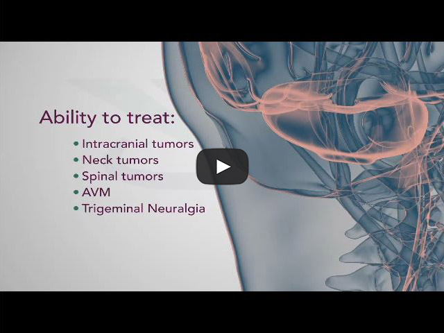
CyberKnife explained by Neurosurgeons
CyberKnife vs. Gamma Knife
The CyberKnife System and Gamma Knife are two of the treatment options for acoustic neuroma. Find out more about the differences between CyberKnife and Gamma Knife here.
What is an Acoustic Neuroma?
An acoustic neuroma, also known as a vestibular schwannoma, is a non-cancerous growth on the eighth cranial nerve, which leads from the brain to the inner ear. This nerve has two distinct parts: one associated with transmitting sound and the other with sending balance information to the brain from the inner ear. The eighth nerve lies adjacent to the facial (seventh cranial) nerve, which provides motion to facial muscles, as they pass through a bony canal called the internal auditory canal. It is at this location where acoustic neuromas originate from the sheath surrounding the eighth nerve.
Acoustic neuromas usually grow slowly over a period of years. They expand in size at their site of origin, and can ultimately displace normal brain tissue. The brain is not invaded by the tumor, but the tumor pushes the brain as it enlarges. The slowly enlarging tumor protrudes from the internal auditory canal into an area behind the temporal bone called the cerebellopontine angle. Larger tumors can press on another nerve in the area (the trigeminal nerve), which produces facial sensation. Vital functions to sustain life can be threatened when large tumors cause severe pressure on the brain stem and cerebellum.
Identifying the Acoustic Neuroma
Advances in medicine have made possible to identify small acoustic neuromas, which are still confined to the internal auditory canal. Routine auditory tests may reveal a loss of hearing and an audiogram should be performed to effectively evaluate hearing in both ears. A loss in one ear should prompt a magnetic resonance imaging (MRI) test.
An MRI with contrast is the preferred diagnostic test for identifying acoustic neuromas. This technique can identify tumors measuring only a few millimeters in diameter.
An auditory brain stem response test (a.k.a. ABR, BAER, or BSER) may be done in some cases. This test provides information on the passage of an electrical impulse along the circuit from the inner ear to the brainstem pathways. An acoustic neuroma can interfere with the passage of this electrical impulse through the hearing nerve at the site of tumor growth in the internal auditory canal, even when the hearing is still essentially normal. This implies the possible diagnosis of an acoustic neuroma when the test result is abnormal. An abnormal auditory brainstem response test should be followed by an MRI.
When an MRI is not available or cannot be performed, a computerized tomography (CT) scan with contrast is suggested for patients in whom an acoustic neuroma is suspected. The combination of CT scan and audiogram approach the reliability of an MRI in making the diagnosis of acoustic neuroma.
Symptoms
Early symptoms are easily overlooked, thus making diagnosis a challenge. In 90 percent of patients with an acoustic neuroma, the first symptom is a reduction in hearing in one ear, often accompanied by ringing in the ear called tinnitus. The loss of hearing is usually subtle and worsens slowly, although occasionally a sudden loss of hearing can occur. There may be a feeling of fullness in the affected ear. These early symptoms are sometimes mistaken for normal changes of aging, or attributed to noise exposure earlier in life, which delays diagnosis.
Since the balance portion of the eighth nerve is where the tumor arises, unsteadiness and balance problems or even vertigo (the feeling like the world is spinning), may occur as the tumor grows. The remainder of the balance system sometimes compensates for this loss, and, in some cases, no balance issues will be noticed. Larger tumors can press on the trigeminal nerve, causing constant or intermittent facial numbness and tingling. Growth of the tumor can put pressure on the brain, resulting in headaches, clumsy gait and mental confusion. These symptoms can be life-threatening complications, which require immediate treatment.
Even though the facial nerve may be compressed by the tumor, it is unusual for patients to experience weakness or paralysis of the face from acoustic neuromas. However, this could occur either in the short- or long-term.
Treatment Options
There are three treatment options available to a patient:
- Observation
- Microsurgical removal
- Radiation (also called radiosurgery or radiotherapy)
Choosing the best treatment is a decision that must be made by both the patient and the physician after careful review of the size of the tumor, its location, as well as the patient's age and physical health.
Observation - Watch and Wait
Acoustic neuromas may be discovered when the tumor is small and symptoms are minor or incidentally when an MRI is performed to evaluate another condition. Since acoustic neuromas are benign tumors and produce symptoms by slowly applying pressure on surrounding nerves, careful observation over a period of time may be appropriate for some patients. When a small tumor is discovered in an older patient, observation to determine the tumor’s growth may be indicated if symptoms are minimal or not present. There is now good evidence from large observational studies that suggest many small tumors in older individuals do not grow, and will not require intervention. Doctors may recommend that MRI scans are performed periodically to keep an eye on the size of the tumor.
Microsurgical Tumor Removal
There are three approaches to surgery for acoustic neuromas:
- Subtotal Removal: Only a portion of the tumor is removed when attempting to remove the entire tumor would risk the patient’s life or neurological function. Periodic MRI studies are important to follow the potential growth of a tumor following this procedure.
- Near Total Tumor Removal: This approach is used by experienced centers when small areas of the tumor are so adherent to the facial nerve that total removal would result in facial weakness. The piece left is generally less than 1 percent of the full tumor and poses a very small risk of regrowth Periodic MRI studies are important to follow the potential growth of a tumor following this procedure.
- Total Tumor Removal: Many tumors can be entirely removed by surgery. Microsurgical techniques and instruments, along with the operating microscope, have greatly reduced the surgical risks of total tumor removal. Preservation of the facial nerve to prevent permanent facial paralysis is the primary task for the experienced acoustic neuroma surgeon. Preservation of hearing is an important goal for patients who present with functional hearing. Both facial nerve function and hearing is electrically monitored during surgery. This is a valuable aid for the surgeon while the tumor is being removed.
Radiation
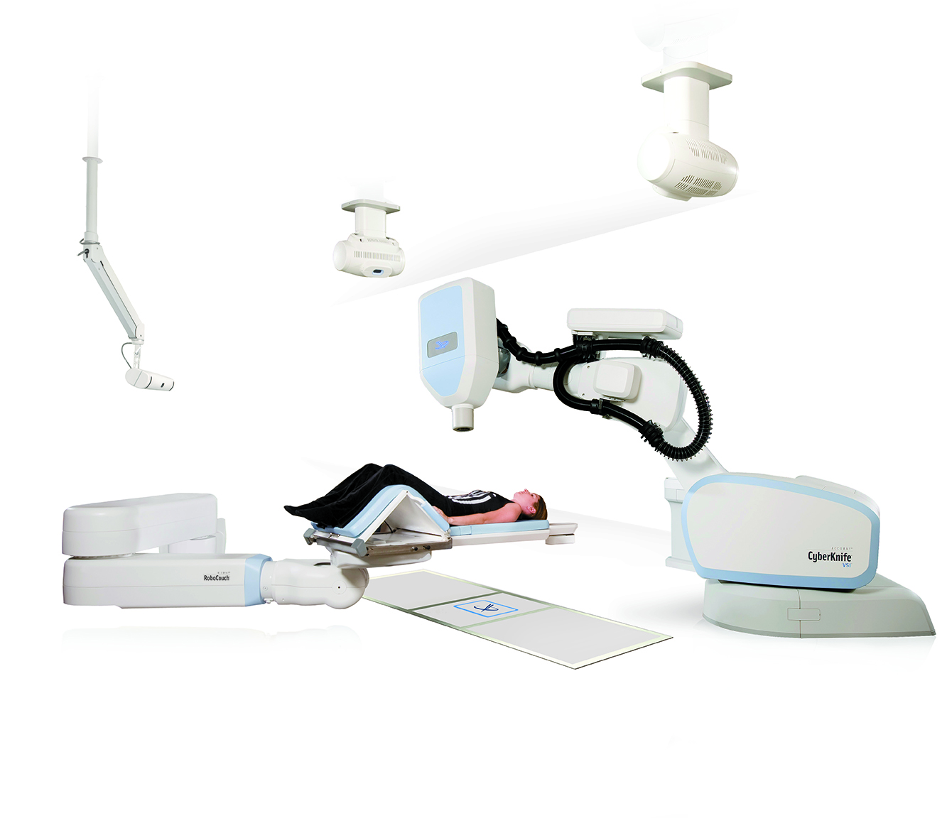
Another treatment option for an acoustic neuroma is radiation, which can be delivered as single-fraction stereotactic radiosurgery (SRS) or as multi-session fractionated stereotactic radiotherapy (FSR). Both techniques are performed in the outpatient setting, not requiring general anesthesia or a hospital stay. The purpose of these techniques is to stop tumor growth and/or cause the tumor to die.
In single-dose treatments, many hundreds of small beams of radiation are aimed at the tumor from various angles around the head, which results in a high dose of radiation to the tumor and very little to any surrounding brain structures. This treatment has had high success rates. Facial weakness or numbness, in the hands of experienced radiation physicians, occurs in only a small percent of cases. Hearing can be preserved in some cases, with a slightly greater opportunity with FSR.
The multi-dose FSR treatment delivers smaller doses of radiation over a period of time, requiring the patient to return to the treatment location on a daily basis, from three to 30 times, generally over several weeks. Each visit lasts a few minutes and most patients are free to go about their daily business before and after each treatment session.
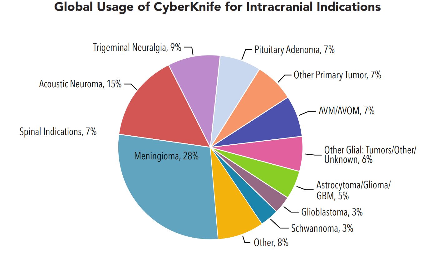
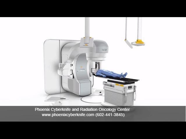
Follow-up after SRS and FSR typically involves an MRI scan and an audiogram at six months, one year, then annually for several years, to ensure the tumor does not start to grow again. Several types of machines deliver focused radiation treatment suitable for treating acoustic neuromas, such as Gamma Knife® and linear accelerator (linac), such as CyberKnife® System, Novalis® TrueBeam STx and Trilogy®.
Patient Education

CyberKnife - Brain, Head & Neck Cancer

TrueBeam STx - Brain, Head & Neck Cancer
TrueBeam STx - Animation
CyberKnife explained by Neurosurgeons
Patient Testimonials

Richard 's CyberKnife Treatment Trigeminal Neuralgia

CyberKnife Treatment Acoustic Neuroma (AN)
CyberKnife explained by Neurosurgeons
CyberKnife vs. Gamma Knife
The CyberKnife System and Gamma Knife are two of the treatment options for acoustic neuroma. Find out more about the differences between CyberKnife and Gamma Knife here.
What is an Acoustic Neuroma?
An acoustic neuroma, also known as a vestibular schwannoma, is a non-cancerous growth on the eighth cranial nerve, which leads from the brain to the inner ear. This nerve has two distinct parts: one associated with transmitting sound and the other with sending balance information to the brain from the inner ear. The eighth nerve lies adjacent to the facial (seventh cranial) nerve, which provides motion to facial muscles, as they pass through a bony canal called the internal auditory canal. It is at this location where acoustic neuromas originate from the sheath surrounding the eighth nerve.
Acoustic neuromas usually grow slowly over a period of years. They expand in size at their site of origin, and can ultimately displace normal brain tissue. The brain is not invaded by the tumor, but the tumor pushes the brain as it enlarges. The slowly enlarging tumor protrudes from the internal auditory canal into an area behind the temporal bone called the cerebellopontine angle. Larger tumors can press on another nerve in the area (the trigeminal nerve), which produces facial sensation. Vital functions to sustain life can be threatened when large tumors cause severe pressure on the brain stem and cerebellum.
CyberKnife explained by Neurosurgeons
Identifying the Acoustic Neuroma
Advances in medicine have made possible to identify small acoustic neuromas, which are still confined to the internal auditory canal. Routine auditory tests may reveal a loss of hearing and an audiogram should be performed to effectively evaluate hearing in both ears. A loss in one ear should prompt a magnetic resonance imaging (MRI) test.
An MRI with contrast is the preferred diagnostic test for identifying acoustic neuromas. This technique can identify tumors measuring only a few millimeters in diameter.
An auditory brain stem response test (a.k.a. ABR, BAER, or BSER) may be done in some cases. This test provides information on the passage of an electrical impulse along the circuit from the inner ear to the brainstem pathways. An acoustic neuroma can interfere with the passage of this electrical impulse through the hearing nerve at the site of tumor growth in the internal auditory canal, even when the hearing is still essentially normal. This implies the possible diagnosis of an acoustic neuroma when the test result is abnormal. An abnormal auditory brainstem response test should be followed by an MRI.
When an MRI is not available or cannot be performed, a computerized tomography (CT) scan with contrast is suggested for patients in whom an acoustic neuroma is suspected. The combination of CT scan and audiogram approach the reliability of an MRI in making the diagnosis of acoustic neuroma.
Symptoms
Early symptoms are easily overlooked, thus making diagnosis a challenge. In 90 percent of patients with an acoustic neuroma, the first symptom is a reduction in hearing in one ear, often accompanied by ringing in the ear called tinnitus. The loss of hearing is usually subtle and worsens slowly, although occasionally a sudden loss of hearing can occur. There may be a feeling of fullness in the affected ear. These early symptoms are sometimes mistaken for normal changes of aging, or attributed to noise exposure earlier in life, which delays diagnosis.
Since the balance portion of the eighth nerve is where the tumor arises, unsteadiness and balance problems or even vertigo (the feeling like the world is spinning), may occur as the tumor grows. The remainder of the balance system sometimes compensates for this loss, and, in some cases, no balance issues will be noticed. Larger tumors can press on the trigeminal nerve, causing constant or intermittent facial numbness and tingling. Growth of the tumor can put pressure on the brain, resulting in headaches, clumsy gait and mental confusion. These symptoms can be life-threatening complications, which require immediate treatment.
Even though the facial nerve may be compressed by the tumor, it is unusual for patients to experience weakness or paralysis of the face from acoustic neuromas. However, this could occur either in the short- or long-term.
Treatment Options
There are three treatment options available to a patient:
- Observation
- Microsurgical removal
- Radiation (also called radiosurgery or radiotherapy)
Choosing the best treatment is a decision that must be made by both the patient and the physician after careful review of the size of the tumor, its location, as well as the patient's age and physical health.
Observation - Watch and Wait
Acoustic neuromas may be discovered when the tumor is small and symptoms are minor or incidentally when an MRI is performed to evaluate another condition. Since acoustic neuromas are benign tumors and produce symptoms by slowly applying pressure on surrounding nerves, careful observation over a period of time may be appropriate for some patients. When a small tumor is discovered in an older patient, observation to determine the tumor’s growth may be indicated if symptoms are minimal or not present. There is now good evidence from large observational studies that suggest many small tumors in older individuals do not grow, and will not require intervention. Doctors may recommend that MRI scans are performed periodically to keep an eye on the size of the tumor.
Microsurgical Tumor Removal
There are three approaches to surgery for acoustic neuromas:
- Subtotal Removal: Only a portion of the tumor is removed when attempting to remove the entire tumor would risk the patient’s life or neurological function. Periodic MRI studies are important to follow the potential growth of a tumor following this procedure.
- Near Total Tumor Removal: This approach is used by experienced centers when small areas of the tumor are so adherent to the facial nerve that total removal would result in facial weakness. The piece left is generally less than 1 percent of the full tumor and poses a very small risk of regrowth Periodic MRI studies are important to follow the potential growth of a tumor following this procedure.
- Total Tumor Removal: Many tumors can be entirely removed by surgery. Microsurgical techniques and instruments, along with the operating microscope, have greatly reduced the surgical risks of total tumor removal. Preservation of the facial nerve to prevent permanent facial paralysis is the primary task for the experienced acoustic neuroma surgeon. Preservation of hearing is an important goal for patients who present with functional hearing. Both facial nerve function and hearing is electrically monitored during surgery. This is a valuable aid for the surgeon while the tumor is being removed.
Radiation
Another treatment option for an acoustic neuroma is radiation, which can be delivered as single-fraction stereotactic radiosurgery (SRS) or as multi-session fractionated stereotactic radiotherapy (FSR). Both techniques are performed in the outpatient setting, not requiring general anesthesia or a hospital stay. The purpose of these techniques is to stop tumor growth and/or cause the tumor to die.
In single-dose treatments, many hundreds of small beams of radiation are aimed at the tumor from various angles around the head, which results in a high dose of radiation to the tumor and very little to any surrounding brain structures. This treatment has had high success rates. Facial weakness or numbness, in the hands of experienced radiation physicians, occurs in only a small percent of cases. Hearing can be preserved in some cases, with a slightly greater opportunity with FSR.
The multi-dose FSR treatment delivers smaller doses of radiation over a period of time, requiring the patient to return to the treatment location on a daily basis, from three to 30 times, generally over several weeks. Each visit lasts a few minutes and most patients are free to go about their daily business before and after each treatment session.
Follow-up after SRS and FSR typically involves an MRI scan and an audiogram at six months, one year, then annually for several years, to ensure the tumor does not start to grow again. Several types of machines deliver focused radiation treatment suitable for treating acoustic neuromas, such as Gamma Knife® and linear accelerator (linac), such as CyberKnife® System, Novalis® TrueBeam STx and Trilogy®.